Design standards for imaging areas are changing
 |
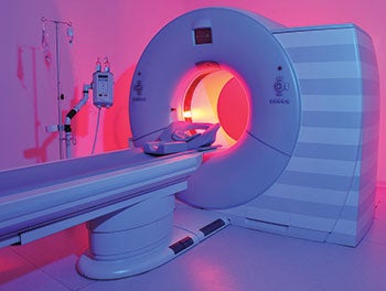
| PHOTO BY LEVENT KONUK/THINKSTOCK Minimum CT clearances have been expanded in the 2014 Guidelines. |
Imaging services account for a disproportionate amount of capital costs when it comes to new hospital construction. It has been estimated that specialty construction, capital equipment costs and service agreements for imaging services can result in up to 50 percent of total project costs for new hospital construction.
Because of the project costs, the complexity of siting advanced imaging modalities and the delays and changes that can be required, it is essential that more foundational elements such as code requirements do not add unwelcome surprises.
The 2014 edition of the Facility Guidelines Institute’s (FGI’s) Guidelines for Design and Construction of Hospitals and Outpatient Facilities offers some notable changes from the 2010 edition with respect to imaging services, and it is important for designers, engineers, facility and equipment planners to familiarize themselves with them.
Beyond the department
For a start, the section title for imaging has been switched from “Diagnostic Imaging Services” to “Imaging Services.” The removal of “diagnostic” from the section title acknowledges that radiology is no longer strictly a department — a collection of rooms around which can be drawn a defined border. Rather, imaging occurs in locations throughout many enterprises and not strictly for diagnostic purposes.
For instance, imaging increasingly is used to inform and direct interventions: from image-guided biopsies via fluoroscopy, computed tomography (CT) or magnetic resonance imaging (MRI) to image-guided surgeries. While the 2014 Guidelines preserves a separate section for “Interventional Imaging,” this change shows that the current edition of the Guidelines more closely reflects contemporary practices versus an either/or approach to diagnostic and interventional imaging services.
Shielded alcoves no longer have a prescriptive distance from exposure control to the edge of the shielded partition wall for all ionizing radiation-emitting modalities, such as X-ray, fluoroscopy and CT. Instead, it is required that the size be suitable to “minimize radiation exposure of technologists and others.” Similarly, the shielded view window now must meet the performance requirement of providing a full view of the exam and procedure table, even when tilted.
As part of recognizing that imaging services now frequently support intervention, the finishes used in patient care areas where imaging services are provided must be selected to conform to the maximum level of interventional care used in each room. Image-guided biopsy, for example, may be comparable to a special procedures room and may require finishes that meet an equivalent level of performance for cleanability and infection control.
Interventional imaging
Throughout the 2014 Guidelines imaging sections, there is repeated reference to meeting the minimum siting conditions from the equipment manufacturers. However, while manufacturer technical minimums are essential, they frequently are not common between vendors or equipment models because of the variability in both equipment and clinical applications.
Because of their often-similar equipment and space configurations, the Guidelines allows for the combination of facilities for interventional imaging and interventional cardiology when it doesn’t conflict with local jurisdictional requirements. Both in terms of general considerations and specific design requirements and recommendations, the 2014 Guidelines greatly increases guidance for interventional procedure preparation and recovery areas.
Radiation shielding requirements for interventional imaging largely mirror those labeled “Diagnostic Imaging” in the 2010 edition, although with an increased emphasis on provision for personal protective equipment. And while control rooms are required, an exception is provided for dedicated electrophysiology procedure rooms, provided the omission is approved by a radiation physicist and appropriate personal radiation protection is provided.
While support areas for imaging services are not individually required, the 2014 Guidelines urges planners to consider the needs for each, and to design accordingly. Changes in the requirements for these recommended areas include emphases on staff work and documentation areas, patient consultation areas, point-of-care lab work or contrast preparation areas, image management systems and reading spaces, and medication safety zones, as appropriate to the services being provided.
The 2014 Guidelines also provides for in-unit processing of ultrasound probes with equipment and operational requirements for probe-cleaning facilities. As the last of conventional film imaging equipment is all but retired, the requirement for “film processing” adjacency also has been removed from the 2014 Guidelines.
Various modalities
A closer examination of changes for various imaging modalities reflects the technical and safety considerations brought to bear in the 2014 Guidelines:
Computed tomography. Of particular note is the increase in minimum clearances around the CT scanner gantry and table. Minimums have been bumped up from 3 feet to 4 feet on all sides of the gantry and table in the 2014 edition. This increase is due, at least in part, to the higher patient acuities and greater space needs for emergent and trauma care that contemporary imaging supports.
Another change from the 2010 edition is that the technologist at the operator’s console must be able to see the patient and the part of the anatomy being scanned. Space constraints in retrofit situations sometimes have meant it is easier to have layouts where the operator’s view is perpendicular to the CT scanner patient table. If this arrangement compromises the technologist’s ability to see the part of the patient in the center of the scanner, it is no longer permitted.
And while it has been seen in practice for many years, the contemporary edition includes language that expressly permits shared control rooms to serve more than one CT scanner room.
X-ray. While the 2014 edition makes strides toward recognizing the continuum of use for all imaging, X-ray modalities still are identified as “Diagnostic Radiography Facilities,” suggesting the transition in the text of the Guidelines remains a work in progress. And just as the title of this section has not changed, the planning and design criteria also are fundamentally unchanged from the prior edition, with one notable exception.
Mammography is no longer a placeholder as it was in 2010. The new criteria include visual privacy, a hand-washing station in the procedure room and changing rooms that are immediately accessible, although these changing rooms may be shared with other functions.
Magnetic resonance imaging. In parallel with CT, minimum clearances of 4 feet have been added around the MRI gantry and patient table. Additionally, the door swing should not encroach on MRI clearances.
While it has always been smart design practice, the Guidelines now requires that MRI equipment siting and site planning take into account potential electromagnetic conflicts between MRI and other equipment. This includes external objects that potentially may interfere with the MRI’s operation, such as elevators, fans, transformers, switchgear and power lines, as well as the MRI’s potential interference with other equipment in the building, such as CT scanners, single-photon emission CT/positron-emission tomography scanners and calibration equipment.
While the FGI Guidelines has referenced the four-zone model of screening, access control and supervision for MRI since the 2010 edition, this has been expanded in 2014 to require design accommodation for containment of non-MRI-safe objects brought to the MRI suite, such as portable oxygen cylinders outside the controlled access zone.
For superconducting MRI systems, cryogen-protection designs should include three elements: venting, emergency exhaust and pressure relief systems. These are to be designed according to the equipment manufacturer’s specifications.
Unlike other areas of patient contact or interventional care for imaging services, the 2014 Guidelines states that hand-washing stations are not required in the MRI scanner room but may be located directly accessible to it. Individual sites’ infection control policies, however, are likely to supersede these FGI requirements.
When a radio-frequency-shielded MRI scanner room door swings outward into a control room or area, the door may not block the view from the console. And, similar to CT, the 2014 Guidelines includes language that explicitly permits shared control rooms that serve more than one MRI scanner room.
MRI suites must have a space identified for code or resuscitation activities located near the control room but beyond the 5-gauss magnetic field extents of the MRI scanner.
If providing interventional services within the MRI suite, sites must provide a pre-procedure preparation and recovery area that complies with the specific provisions in the “Interventional Imaging” section.
In a move parallel to the design criteria to minimize interference between the MRI scanner and other equipment in the building, the Guidelines now requires the MRI scanner room floor design to take into account the thresholds for ferrous materials for the MRI scanner, or shim tolerances, to help protect image quality.
The 2014 Guidelines also adds a specific reference for sound isolation performance, requiring that designs address acoustic control measures between the MRI scanner room and adjacent areas.
Specific changes
While not intended to serve as a substitute for the 2014 FGI Guidelines, this summary of the more significant changes for sections 2.2–3.4, Imaging Services, and 2.2–3.5, Interventional Imaging, should help to identify many of the specific changes from the prior edition for these two sections.
Changes in imaging technology and clinical practice continue to accelerate and may require special accommodation even before the next iteration of the Guidelines is published. One concrete example of this is the inclusion of a new section title in the 2014 Guidelines for “Interventional and Intraoperative MRI (I-MRI) Facilities,” which provides no specific requirements, but telegraphs the expectation of future standards.
Given the complexities and costs associated with imaging facilities, seemingly modest changes could wind up having tens or even hundreds of thousands of dollars of cost consequences if not discovered early in the process.
The rapidly advancing technical and clinical demands on imaging services will continue to require higher and higher levels of expertise from designers and the design elements from the most recent FGI Guidelines are an essential start.
is senior vice president with Radiology-Planning of Kansas City, Mo., and principal of Gilk Radiology Consulting. He also has served as a special imaging consultant to the 2010 and 2014 editions of the Facility Guidelines Institute’s Guidelines for Design and Construction of Hospitals and Outpatient Facilities. He can be reached at tgilk@RAD-Planning.com.
Facility planning principles for imaging services
The following encapsulates some of the major planning principles for imaging facilities from the 2014 edition of the Facility Guidelines Institute’s (FGI’s) Guidelines for Design and Construction of Hospitals and Outpatient Facilities.
These may be helpful as part of a standard planning and design review for radiology and nuclear medicine facilities:
• To preserve flexibility, facilities professionals should evaluate every new imaging modality siting against the facility’s infection control standards for interventional care. This evaluation should consider finishes, ventilation and hand-washing requirements. Future demands over the projected lifespan of the imaging equipment should be anticipated to minimize risks of future patient care-interrupting modifications to the site.
• Particularly for reuse and upgrades of existing imaging scanning rooms, facilities professionals should verify that the dimensions of the room will accommodate increased clearance requirements. Rooms designed to the minimum requirements of prior editions of the FGI Guidelines or other design standards and codes may not meet the 2014 standards.
• Patient visibility during imaging is receiving greater attention. When planning new imaging equipment installations, facilities professionals should make certain that the technologist can clearly see the patient and the anatomy being imaged from the operator’s console.
• In the absence of specific state, federal or accreditation standards, the 2014 Guidelines is reaffirming itself as the standard-bearer for physical magnetic resonance imaging (MRI) safety protections. As a general guideline for MRI equipment siting, facilities professionals should make sure to adhere to both the equipment manufacturer’s safety guidance and the most recent MRI safety guidance from the American College of Radiology.
• When planning facilities for new imaging equipment, it is advisable to compare the physical siting requirements of competing systems so that an individual room design can accommodate the minimum requirements of different vendors.
These planning principles represent larger themes found in the FGI Guidelines. Individual facilities likely will have additional design standards imposed by enterprise- or modality-level accreditation providers and may have to meet other requirements from local or state authorities as well.


